
The brainstem is not a single “control center,” but a major integration region linking the brain with the spinal cord and helping coordinate breathing, cardiovascular regulation, sleep and arousal, swallowing, vomiting, eye movements, hearing and balance functions, facial movement and sensation, and multiple autonomic processes. (Benarroch, 2017; McCorry, 2007)
This article examines how brainstem-mediated autonomic function, cranial nerve pathways, vestibular function, sensory-motor organization, defensive reflexes, and regulation can come together in the clinical picture.
The brainstem is an important site of autonomic integration. It participates in cardiovascular regulation, respiration, swallowing, arousal, sleep, vestibular processing, eye movements, visceral signaling, and cranial nerve function. When these systems become poorly coordinated, patients may present with symptoms that appear unrelated: orthostatic intolerance, palpitations, nausea, digestive slowing, dizziness, visual strain, sound sensitivity, facial tension, altered voice, swallowing difficulty, disrupted sleep, poor balance, motor inhibition, and persistent high-tone bracing. In our clinical work, this is the presentation we see most often in Long COVID.
The purpose of this spoke is not to assume that these findings arise from one cause or to replace medical evaluation. Cranial nerve symptoms, abnormal neurological signs, severe autonomic symptoms, and progressive functional loss can result from many neurological, cardiovascular, endocrine, autoimmune, infectious, structural, medication-related, sleep-related, and post-infectious conditions. The purpose is to recognize when these findings form a broader neurological-autonomic pattern, to assess how that pattern is affecting function, and to determine where treatment can safely begin.
Why the Brainstem Matters
The autonomic nervous system does not operate as a single nerve or a single pathway. It is a distributed regulatory network involving the brain, hypothalamus, limbic system, brainstem, spinal cord, peripheral nerves, autonomic ganglia, cardiovascular system, lungs, gut, immune system, endocrine system, and sensory systems. The brainstem is one of the central locations where incoming signals from the body are integrated with outgoing autonomic instructions.
The medulla and pons help regulate breathing, heart rate, blood pressure, swallowing, balance, wakefulness, and cranial nerve function. When that regulation breaks down, patients may show autonomic symptoms, changes in cranial nerve function, sensory or motor changes, altered consciousness, or problems with breathing. None of these symptoms alone proves brainstem disease. The same symptoms can come from many other causes and may require medical evaluation. (Martín-Gallego et al., 2017; McCorry, 2007).
For this reason, the clinical assessment begins with pattern recognition rather than conclusion.
The clinician asks:
- Which functions are affected?
- Did the symptoms begin suddenly, gradually, after illness, after surgery, after injury, or during a period of sustained overload?
- Are symptoms stable, intermittent, progressive, positional, exertional, sensory-triggered, or linked to sleep?
- Which findings are new, asymmetrical, severe, or worsening?
- What medical assessment has already occurred?
- Which systems appear to change together?
A patient may experience dizziness, nausea, visual sensitivity, vocal fatigue, jaw clenching, restricted neck movement, poor sleep, digestive slowing, and a rapid heart rate upon standing. None of those findings alone establishes a single diagnosis. Together, however, they identify a clinical cluster that deserves a more integrated neurological-autonomic assessment.
Cranial Nerve Function
The cranial nerves carry motor, sensory, and autonomic information involving the face, eyes, ears, throat, tongue, jaw, neck, heart, lungs, and digestive tract. (Miller et al., 2009; Bajaj et al., 2014; McCorry, 2007)
Cranial nerves provide a practical window into functions that involve the face, jaw, eyes, ears, palate, tongue, swallowing, voice, vestibular system, and parasympathetic regulation. In a patient with multisystem dysautonomia, a careful history of these functions may reveal patterns that are otherwise missed when symptoms are divided among dental, gastrointestinal, neurological, otolaryngological, ophthalmological, psychiatric, and cardiovascular categories.
Facial, Jaw, Palatal, and Tongue Function
Clinical observations may include facial asymmetry, changes in facial expression, chronic jaw clenching, temporomandibular tension, difficulty opening the mouth, tongue tension, altered tongue movement, palatal changes, or difficulty coordinating chewing and swallowing.
These findings can occur with pain, dental conditions, temporomandibular disorders, cervical dysfunction, medication effects, peripheral nerve disorders, central neurological disease, movement disorders, sleep disturbance, trauma-related bracing, and many other conditions. They should not be interpreted automatically as signs of autonomic dysregulation or trauma physiology.
Within the broader autonomic pattern, however, chronic jaw, tongue, throat, and upper-cervical tension can be clinically relevant. These regions participate in breathing, swallowing, vocalization, facial expression, orienting, social engagement, threat detection, and protective bracing. A patient who chronically holds the jaw, tongue, neck, and diaphragm in a defensive configuration may have difficulty breathing freely, swallowing comfortably, speaking with ease, tolerating sensory input, or shifting out of a high-alert state.
The clinical question is not, “Is this trauma or is it neurological?” The question is, “What is this patient’s pattern of motor, sensory, autonomic, structural, and medical contributors and what requires further evaluation?”
Voice, Swallowing, and Airway Protection
Voice and swallowing are especially important because they involve safety, nutrition, hydration, respiration, airway protection, communication, and vagal/brainstem-connected functions. Relevant symptoms include:
- Hoarseness or loss of vocal endurance.
- Frequent throat clearing.
- A sensation of throat tightness.
- Difficulty initiating a swallow.
- Coughing or choking with food or liquids.
- Food sticking in the throat.
- Changes in speech articulation.
- Recurrent aspiration or chest infections.
- Unexplained weight loss related to eating difficulty.
- Severe reflux, nausea, or vomiting.
Some clients describe a throat that “closes” during stress, panic, grief, conflict, sensory overload, or periods of intense fatigue. That experience can be clinically meaningful and may coexist with muscular guarding, altered breathing, reflux, laryngeal hypersensitivity, autonomic activation, or conditioned protective responses. It should not, however, be assumed to be solely emotional or functional. Persistent or progressive dysphagia, choking, aspiration, unexplained weight loss, voice change, or new speech disturbance requires medical evaluation.
Ocular-Motor and Vestibular Function
The eyes and vestibular system are continuously involved in orientation, balance, posture, motion tolerance, spatial safety, and autonomic regulation. Patients with autonomic dysregulation may report:
- Dizziness or lightheadedness.
- Motion sensitivity.
- Nausea in visually busy environments.
- Difficulty tracking moving objects.
- Visual fatigue.
- Difficulty shifting focus from near to far.
- Poor tolerance for screens, fluorescent lights, crowds, or driving.
- Imbalance or unsteadiness.
- A sense of disorientation in open spaces, stores, stairs, or moving environments.
- Symptoms that intensify with fatigue, upright posture, pain, infection, or sleep loss.
Vestibular symptoms can amplify autonomic dysregulation because loss of orientation is itself threatening to the nervous system. A person who becomes dizzy, nauseated, or disorganized in visually complex environments may begin to brace, restrict movement, avoid turning the head, hold the breath, withdraw from activity, or remain hypervigilant. These adaptations can reduce immediate discomfort while gradually narrowing the person’s functional range.
But dizziness and visual symptoms have broad differentials. They may relate to vestibular migraine, benign positional vertigo, inner-ear disease, concussion, medication effects, visual disorders, cardiovascular disease, anemia, dehydration, orthostatic intolerance, neurological disease, anxiety, or sleep disturbance. New double vision, persistent nystagmus, abrupt severe headache, focal weakness, facial droop, speech disturbance, sudden hearing loss, or inability to walk safely requires urgent medical assessment.
Auditory Sensitivity and Startle
Sound sensitivity, exaggerated startle, difficulty filtering background noise, hyperacusis, tinnitus, and distress in noisy environments are common complaints among patients with chronic dysregulation. These symptoms may reflect auditory conditions, migraine, sensory-processing differences, concussion, medication effects, sleep deprivation, autonomic arousal, chronic pain, or threat-related vigilance.
In the autonomic framework, sound sensitivity may be understood as a nervous system that is allocating too much attention to possible threat. The person does not merely “dislike noise.” The nervous system may treat ordinary input as demanding, invasive, unpredictable, or difficult to filter. This can create a cycle in which sensory overload increases arousal, arousal reduces filtering capacity, and reduced filtering capacity makes the environment feel even more dangerous.
The treatment implication is not forced exposure or telling a patient to tolerate more than their system can manage. It is gradual restoration of capacity through pacing, environmental modification, sleep support, medical assessment when needed, carefully dosed sensory input, breathing and postural work, movement tolerance, and improved ability to recover after activation.
Primitive Reflexes and Defensive Motor Patterns
Primitive reflexes are automatic motor responses that are most visible during early development. Their clinical interpretation in adults requires caution. Some neurological signs traditionally described as primitive reflexes can be relevant in formal neurological examination, especially when new or clearly pathological. Other reflex-integration frameworks use the language of retained, reactivated, or unintegrated reflexes to describe observable patterns of startle, bracing, withdrawal, postural adaptation, movement inhibition, sensory sensitivity, and stress reactivity.
In this clinical model, reflex observations are not treated as standalone proof of a lesion, developmental diagnosis, or trauma mechanism. They are treated as one source of information about how the person organizes protection, movement, attention, and response to sensory input.
Moro and Startle-Related Patterns
A high startle response may appear as sudden shoulder elevation, breath holding, eye widening, rapid muscle recruitment, panic-like activation, flinching, or difficulty recovering after a surprise. It may be intensified by sound, visual motion, touch, interpersonal proximity, pain, fatigue, illness, or sleep deprivation.
Clinically, the relevant question is not whether the patient “has a Moro reflex.” It is whether their startle system is easily recruited and whether they can return to baseline afterward. A person may show a strong startle response and then recover quickly. Another may remain tachycardic, tense, nauseated, dizzy, exhausted, or emotionally overwhelmed for hours. The recovery pattern often tells the clinician more than the startle itself.
Fear, Withdrawal, and Freeze Patterns
Some patients respond to overload not with obvious activation but with inhibition. They may become quiet, immobile, dissociated, cognitively blank, physically rigid, unable to speak, unable to make decisions, unable to initiate movement, or unable to access previously available skills. They may look calm externally while reporting intense internal alarm, dread, pressure, nausea, or exhaustion.
This presentation can resemble depression, dissociation, shutdown, fatigue, executive dysfunction, learned helplessness, pain inhibition, autonomic collapse, or a neurological problem. It may also coexist with any of those conditions. The clinical value of recognizing a freeze-like pattern is that it prevents the practitioner from misreading immobility as lack of motivation, resistance, indifference, or unwillingness.
Spinal Galant and Trunk Reactivity
Patterns described in reflex-integration work as Spinal Galant reactivity may include sensitivity along the paraspinal region, asymmetrical trunk tension, difficulty settling into seated postures, pelvic or hip tension, postural shifting, sleep disruption, and exaggerated reactivity to touch or clothing around the waist. These observations should be interpreted carefully and not treated as diagnostic in isolation.
Within a whole-body assessment, trunk asymmetry and persistent paraspinal reactivity may be relevant because the trunk is central to breathing, gait, pelvic-floor function, spinal rotation, visceral comfort, and the capacity to shift between mobilization and rest. A person who cannot release through the thorax, diaphragm, psoas, lumbar spine, pelvic floor, or cervical region may have reduced access to efficient movement and recovery.
The point is not that a reflex pattern “causes” dysautonomia. The point is that motor organization, sensory reactivity, breathing, posture, and autonomic state continually influence one another.
Autonomic Clues in the Neurological Examination
The neurology becomes important when cranial-nerve-related symptoms occur alongside orthostatic, respiratory, digestive, sleep, motor, and sensory changes. The following domains help organize assessment.
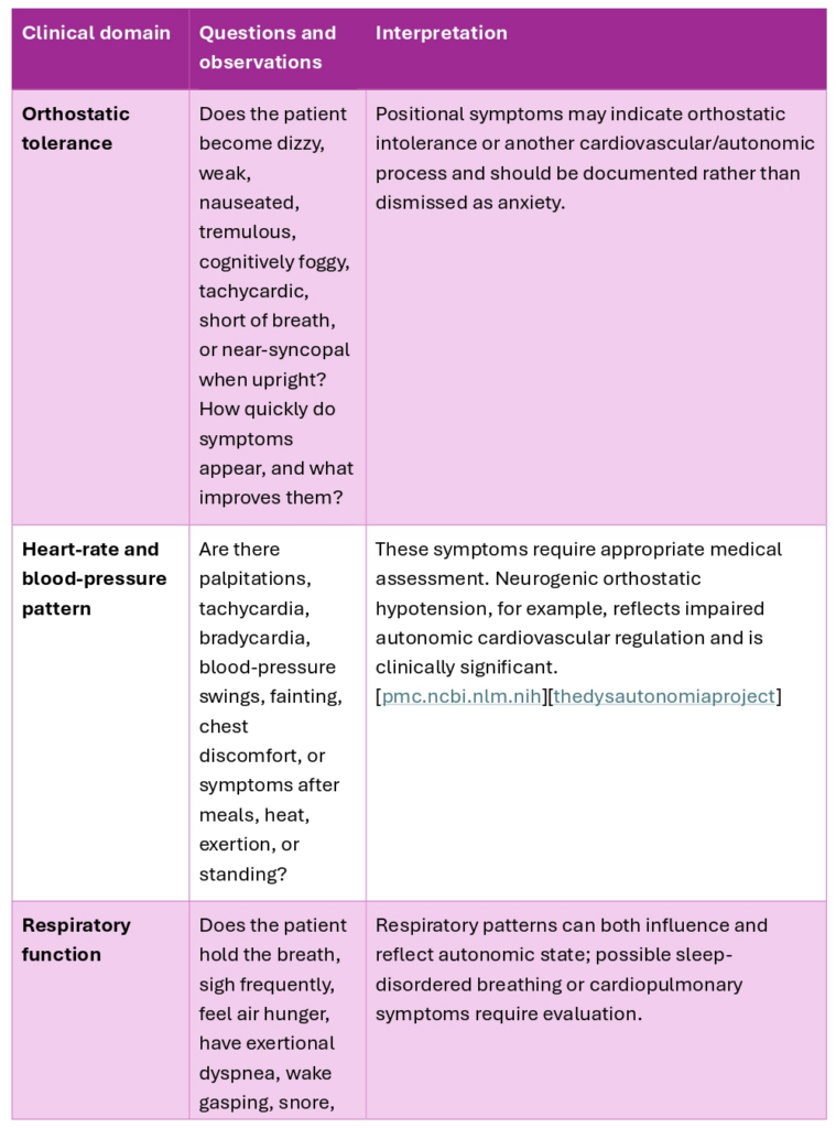
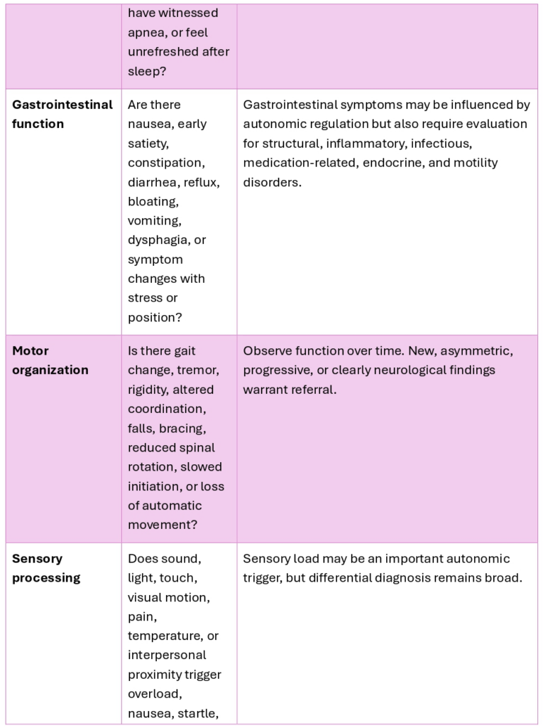
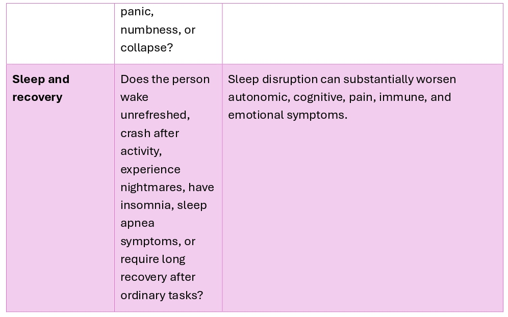
A practical intake question is:
“What happens in your body when you stand, move, eat, speak for a long time, enter a noisy environment, turn your head, try to sleep, become emotionally activated, or push through fatigue?”
That question often reveals whether the patient’s symptoms are primarily positional, exertional, sensory-triggered, digestive, social, sleep-related, pain-linked, emotionally linked, or mixed. It also helps identify the person’s sequence: what comes first, what escalates next, and what finally causes collapse or recovery.
Pattern Differentiation in the Neurological Spoke
The same cranial-nerve, vestibular, sensory, or motor complaint may appear in different autonomic patterns. The clinician therefore asks not only, “What symptom is present?” but also, “What autonomic state is organizing this symptom today?”
Sympathetic Dominance
In sympathetic dominance, the patient often presents with speed, vigilance, tension, over-recruitment, and difficulty downshifting. The neurological expression may include:
- Hyperacusis or sound sensitivity.
- Exaggerated startle.
- Jaw clenching and upper-cervical tension.
- Rapid speech or vocal strain.
- Visual scanning and difficulty resting the eyes.
- Tremor or internal shaking.
- Tension headaches.
- Shallow breathing or breath holding.
- Difficulty sleeping despite exhaustion.
- High muscle tone through the neck, shoulders, psoas, calves, and standing musculature.
The immediate treatment task is usually not emotional excavation. It is to reduce unnecessary load and restore enough safety, orientation, sensory tolerance, breath capacity, postural support, sleep, and pacing that the patient can downshift without collapse.
Mixed Activation and Collapse
In mixed patterns, the patient alternates between mobilization and depletion. They may appear capable, productive, and highly functional during one period, then become cognitively foggy, dizzy, exhausted, nauseated, emotionally overwhelmed, or unable to initiate basic tasks after seemingly modest demand.
The neurological expression may include:
- Variable balance and visual tolerance.
- Fluctuating voice, swallowing comfort, or jaw tension.
- Intermittent tremor, internal agitation, or weakness.
- Changing orthostatic tolerance.
- Alternating insomnia and hypersomnia.
- Sensory overload followed by numbness or withdrawal.
- Periods of intense productivity followed by prolonged crashes.
- Cognitive fluctuation, especially under exertion, time pressure, or emotional demand.
The clinical error is to see the higher-functioning period as proof that the patient is well. In mixed dysautonomia, the apparent “good day” may be driven by overmobilization that cannot be sustained.
Freeze and High-Tone Bracing
In a freeze-dominant pattern, the patient may show reduced movement, restricted rotation, reduced facial expressiveness, shallow respiration, vocal inhibition, cognitive slowing, numbness, immobility, dissociation, and persistent muscular bracing. The person may describe being unable to act despite wanting to act.
The neurological expression may include:
- Restricted diaphragmatic excursion.
- Jaw, tongue, throat, and upper-cervical tension.
- Trunk rigidity and reduced spinal rotation.
- Difficulty initiating movement.
- Visual fixation or difficulty shifting attention.
- Reduced vocal range or a constricted voice.
- Persistent postural holding.
- Numbness, derealization, or dissociation.
- Digestive slowing, nausea, constipation, or reduced appetite.
- Exhaustion that does not resolve fully with rest.
The treatment task is to avoid forcing activation faster than the system can tolerate. Small increases in orientation, breath, movement, sensory choice, postural support, rhythm, voice, and social safety may be more useful than pushing for catharsis, intense exercise, emotional exposure, or insight before the person has enough physiological capacity to remain present.
Medical Red Flags and Referral
A trauma history, chronic stress pattern, reflex-integration finding, fascial restriction, or recognizable autonomic presentation must never be used to explain away serious medical symptoms. The following findings require timely medical assessment, and some may require urgent or emergency evaluation:
- New facial weakness, facial droop, tongue deviation, persistent double vision, new abnormal pupils, sudden hearing loss, or new severe imbalance.
- New, progressive, or persistent dysphagia; choking; aspiration; severe voice change; inability to maintain hydration; or unexplained weight loss.
- Syncope, recurrent near-syncope, chest pain, sustained bradycardia, severe tachycardia, marked blood-pressure instability, or new exertional shortness of breath.
- New weakness, progressive gait deterioration, falls, marked spasticity, loss of coordination, tremor with neurological change, or sensory loss.
- Witnessed apnea, waking gasping, cyanosis, severe daytime sleepiness, morning headaches, or suspected central sleep apnea.
- New bowel or bladder retention, new incontinence with neurological symptoms, saddle numbness, or rapidly changing lower-extremity weakness.
- Persistent vomiting, severe gastroparesis symptoms, dehydration, inability to maintain nutrition, or acute abdominal symptoms.
- Abrupt severe headache, altered consciousness, seizures, stroke-like symptoms, or rapid neurological decline.
The clinician’s role is to document the pattern, recognize when symptoms exceed the scope of body-based care, coordinate with the medical team when appropriate, and avoid reducing potentially serious signs to “dysregulation.”
Treatment Implications
Brainstem issues do not imply that all neurological-autonomic symptoms can be treated through manual therapy, acupuncture, breathing practices, reflex work, somatic coaching, or trauma-informed care. It does suggest that treatment may be more effective when the patient’s autonomic capacity, sensory threshold, motor organization, sleep, breathing, pain, pacing, and medical contributors are considered together.
For some patients, the first intervention is medical stabilization: hydration, medication review, sleep evaluation, cardiology or neurology referral, treatment of anemia or endocrine disease, vestibular assessment, gastrointestinal evaluation, infection workup, management of autoimmune disease, rehabilitation, or physical conditioning adapted to orthostatic tolerance.
For others, carefully paced bottom-up work may help reduce the burden carried by the neurological-autonomic system. The aim is not to force release, create a dramatic emotional response, or prove a trauma theory. The aim is to increase the patient’s ability to remain present in their body without escalating into panic, bracing, dizziness, collapse, pain, sensory overload, or shutdown.
Useful clinical targets may include:
- Improving tolerance for upright posture and graded movement.
- Reducing unnecessary cervical, jaw, diaphragmatic, and trunk bracing.
- Supporting slower, more flexible respiratory patterns without forcing deep breathing.
- Increasing orienting capacity and sensory choice.
- Building tolerance for visual, auditory, vestibular, and interpersonal input in small doses.
- Improving sleep regularity and post-exertional recovery.
- Supporting efficient movement rather than increasing effort.
- Helping the patient recognize early signs of overmobilization before a crash occurs.
- Coordinating treatment with appropriate medical evaluation and rehabilitation.
Stabilization, Patient Agency, and Polyvagal Touch™
The patient who has spent years forcing function through sympathetic overdrive, muscular bracing, dissociation, perfectionism, pain suppression, or repeated crashes may need a treatment plan that begins with stabilization rather than performance. This is especially true for patients whose nervous systems were organized during periods of overwhelming threat in the developmental window. That history may include chronic developmental trauma, but it may also include early medical trauma, such as major surgery, prolonged hospitalization, repeated invasive procedures, cancer treatment, or separation from caregivers during the first year of life.
For an infant or young child, major surgery can represent an annihilating level of threat: the body is overwhelmed, immobilized, invaded, unable to escape, and dependent on others for survival. In the model developed throughout this work, this is the level of threat at which PAG-mediated defensive organization becomes clinically relevant.
Many of these patients remain organized around profound defense and hypervigilance. They may react to the smallest body sensation, a shift in breathing, an unfamiliar touch, a change in position, or a moment of internal quiet as though something dangerous has happened. Their nervous system asks, “What touched me?” The question is an immediate protective response.
This is not resistance, exaggeration, or failure to cooperate. It is a profound state of self-protection. As practitioners, we do not force that defense open. We do not demand relaxation, catharsis, or emotional disclosure before the person has sufficient capacity to remain present. Stabilization begins by respecting the defense, restoring choice, and allowing the patient to control pace, contact, sensation, and participation.
In these patients, therapy without any form of body-based work is often not enough. They are able to discuss their own trauma while remaining protected from the defensive patterns through which that trauma continues to be expressed. Verbal insight is present, but the person may still be unable to feel the body clearly, tolerate internal sensation, receive touch, or remain present when triggered.
This is why stabilization requires more than asking the patient to relax, think differently, or talk about what happened. It requires a safe way to approach the body without overwhelming it. For many patients, mental-health support and gentle, nonintrusive body-based work serve complementary roles to help reconnect the body with emotions.
POLYVAGAL TOUCH™: A Polyvagal Acupuncture® Method
Needle-Free Guided Self-Regulation
Polyvagal Acupuncture® was developed from my own work to restore brainstem regulation after injury and severe dysautonomia. Polyvagal Touch™ is the needle-free form of this work. It can be taught directly to patients, including by Zoom, so that people who are not ready for bodywork, cannot afford ongoing hands-on care, or wish to begin privately can start exploring their own somatic patterns in a structured and tolerable way.
The work is not done to the patient; rather, the patient does the work themselves. Polyvagal Touch™ gives them a way to approach their own body, breath, heart, and sensation without forcing contact, catharsis, or emotional disclosure. The practitioner provides instruction, pacing, consent, observation, and a sufficiently safe relational environment for the patient to notice what happens in the body without immediately needing to defend against it.
Many survivors of developmental trauma live in tonic immobility (Volchan et al., 2011; Kearney & Lanius, 2022; Reynolds et al., 2015). They freeze quickly. The body locks down before tremor, discharge, movement, or emotional processing can begin. When touch is nonintrusive, slow, and chosen by the patient, deep pressure bypasses the brainstem-mediated threat response (Kearney & Lanius, 2022; Reynolds et al., 2015). It gives the patient a direct way to contact the held tissue without having to mobilize, disclose traumatic memory, or force a release. The patient can remain with the pressure, feel the area that holds, and notice what changes.
Polyvagal Touch™ gives patients and practitioners objective markers of change in fascial tone. These changes can be seen and palpated. The patient can learn to recognize when tissue is held, braced, dense, rigid, or guarded, and when it begins to soften, lengthen, and release. The practitioner can observe and palpate these same changes. This gives the patient a concrete guide for recognizing changes in protective holding, rather than relying only on whether they think or feel better.
This matters for patients who have learned not to trust internal sensation. The visible and palpable changes give them something concrete to work with. They can observe what changes with contact, breath, and pacing, and they can stop before the work becomes overwhelming. Over time, this gives the patient a way to recognize their own physiological shifts rather than depending entirely on the practitioner to tell them what is happening.
The goal is not to push the patient into deeper work before they are ready. It is to restore agency: to help the patient begin to trust their own body, recognize changes in protective holding, and develop enough safety and choice to explore more skilled somatic work when their circumstances allow or when they feel moved to do so.
References
Photo: OpenStax, CC BY 4.0 <https://creativecommons.org/licenses/by/4.0>, via Wikimedia Commons
Bajaj, J., et al. (2014). Disorders of the lower cranial nerves. Journal of Neurosciences in Rural Practice.
Benarroch, E. E. (2017). Brainstem and autonomic nervous system dysfunction. Continuum: Lifelong Learning in Neurology, 23(2), 1–20.
Bennet, et al. (2017). Nervous System.
Gibbons, C. H., et al. (2017). The recommendations of a consensus panel for the screening, diagnosis, and treatment of neurogenic orthostatic hypotension and associated supine hypertension. Journal of Neurology, 264, 1567–1582.
Heller, D. P., & Heller, L. (2021). Somatic experiencing—Effectiveness and key factors of a body-oriented trauma therapy: A scoping literature review. European Journal of Psychotraumatology, 12(1), Article 1929023. https://doi.org/10.1080/20008198.2021.1929023
Kearney, B. E., & Lanius, R. A. (2022). The brain-body disconnect: A somatic sensory basis for trauma-related disorders. Frontiers in Neuroscience, 16, Article 1015749. https://doi.org/10.3389/fnins.2022.1015749
Martín-Gallego, A., González-García, L., Carrasco-Brenes, M., Segura-Fernández-Nogueras, M., Delgado-Babiano, A., Ros-Sanjuán, A., Romero-Moreno, L., Domínguez-Páez, M., Dawid-Milner, M. S., & Arráez-Sánchez, M. A. (2017). Brainstem and autonomic nervous system dysfunction: A neurosurgical point of view. Acta Neurochirurgica Supplement, 124, 221–229. https://doi.org/10.1007/978-3-319-39546-3_34pubmed.ncbi.nlm.nih
Miller, R. G., et al. (2009). Disorders of cranial nerves IX and X. Seminars in Neurology.
McCorry, L. K. (2007). Physiology of the autonomic nervous system. American Journal of Pharmaceutical Education, 71(4), Article 78. https://doi.org/10.5688/aj710478pmc.ncbi.nlm.nih
Reynolds, S., Lane, S. J., & Mullen, B. (2015). Brief report—Effects of deep pressure stimulation on physiological arousal. American Journal of Occupational Therapy, 69(3), 6903350010p1–6903350010p5. https://doi.org/10.5014/ajot.2015.015560
Volchan, E., Souza, G. G. L., Franklin, C. M., Norte, C. E., Rocha-Rego, V., Oliveira, J. M., David, I. A., Mendlowicz, M. V., Coutinho, E. S. F., & Fiszman, A. (2011). Is there tonic immobility in humans? Biological evidence from victims of traumatic stress. Biological Psychology, 88(1), 13–19. https://doi.org/10.1016/j.biopsycho.2011.06.002
Walker, S. C., McGlone, F. P., & McGlone, F. (2017). C-tactile afferents: Cutaneous mediators of oxytocin release during affiliative tactile interactions? Neuropeptides, 64, 27–38. https://doi.org/10.1016/j.npep.2017.01.001
Wieling, W., Kaufmann, H., Claydon, V. E., van Wijnen, V. K., Harms, M. P. M., Juraschek, S. P., Thijs, R. D., & colleagues. (2022). Diagnosis and treatment of orthostatic hypotension. The Lancet Neurology, 21(8), 735–746. https://doi.org/10.1016/S1474-4422(22)00169-7
