
The Four Patterns of Dysautonomia
The primary purpose of the autonomic nervous system is survival. Every second it asks one question: Am I safe? (Porges, 2011) The answer determines how blood flows, how muscles contract, how the eyes scan the environment, how food is digested, and ultimately whether the body prepares for defense or recovery. These physiological adjustments occur automatically, long before conscious thought has time to intervene.
When danger is brief, the nervous system recruits a defensive response and then returns to homeostasis once safety is restored. When defensive physiology persists, however, survival becomes the body’s new operating system. Resources shift away from growth, repair, reproduction, digestion, learning, and neuroplasticity toward protection. This shift affects every organ system because every organ system depends on autonomic regulation. (McEwen, 2007; Porges, 2011)
Most people associate the sympathetic nervous system with fight-flight response orstress, but the word stress often obscures this process because it is commonly understood as an emotional or psychological experience. Throughout this synthesis, we will use the word stress to refer to a physiological or emotional state of autonomic defense. Many patients with chronic dysautonomia do not identify as “stressed” because sympathetic activation or freeze has become their normal state. This is particularly common among first responders, military personnel, healthcare workers, working parents and those in support professions. The physiology remains the same whether the patient identifies with the word emotionally.
The autonomic nervous system does not generate an unlimited number of defensive responses. It recruits survival strategies according to the degree of perceived threat. Whether initiated by trauma, major surgery, neurodegenerative disease, autoimmunity, long COVID, or other prolonged illness, chronic autonomic dysregulation organize into four recurring clinical presentations. Western medicine typically classifies these patients according to individual diagnoses. TCM pattern differentiation provides a basis for organizing dysautonomia into clinically useful subtypes to guide treatment.
These four presentations are not separate diseases, and patients often move between them or express features of more than one simultaneously. They are best understood as different expressions of the same dysregulated autonomic system.
The Evolution of a Polyvagal Approach
Porges’s Polyvagal Theory named the hierarchy of autonomic states, ventral vagal, sympathetic, and dorsal vagal, and introduced neuroception, the nervous system’s unconscious read of safety or threat that determines which state takes over. (Porges, 2011). Van der Kolk showed how the body registers and sustains that dorsal vagal freeze internally, long before it becomes visible from the outside. (van der Kolk, 2014). Masgutova’s MNRI work was the first to explicitly map the freeze reflex layer in trauma survivors and establish that this foundation must be addressed before anything downstream can integrate. (Masgutova & Akhmatova, 2011). Levine’s Somatic Experiencing works through thixotropy and the spontaneous tremor discharge of stuck sympathetic activation. (Levine, 2010). Heller’s NARM framework provided the developmental map: where in the sequence the trauma landed determines what organizes downstream. (Heller & LaPierre, 2012). Reich and Lowen showed that the resulting fascial armoring is the body’s structural record of its emotional and limbic history, readable and addressable through the body directly. (Lowen, 1987)
What these approaches have collectively demonstrated is that chronic autonomic dysregulation becomes embodied. (van der Kolk, 2014; Porges, 2011). The nervous system, connective tissue, postural system, and defensive reflexes adapt to chronic threat and continue to express that adaptation long after the threat has passed. (McEwen, 2007; Schleip, 2003).
Dysautonomia, by definition, involves some degree of degraded fight or flight response. Cranial nerve involvement and persistent primitive reflexes can reappear regardless of whether the initiating event was trauma, severe illness, major surgery, long COVID, or neurodegenerative disease. In some patients, the T1 spinal level and stellate ganglion output remain diminished and inhibited, and the nervous system defaults to freeze because active defense is not available. (Porges, 2011) In others, that same circuitry runs in the opposite direction, producing a sympathetic surge that overshoots into chronic hyperarousal rather than shutdown. Which pattern predominates, and when in the nervous system’s development it took hold, shapes the clinical picture and where treatment begins. (Moffitt, 2025)
Sympathetic Dominance (Yang Form)

Sympathetic dominance represents a retained fight or flight response. The body remains poised to flee or engage. Every physiological system prepares for rapid movement, like a sprinter waiting in the blocks.
Sensory information passes through the thalamus before reaching the amygdala, where threat is rapidly assessed. The hypothalamus recruits the sympathetic nervous system and the HPA axis, shifting the body from homeostasis toward active defense. (McEwen, 2007) Speed becomes more important than precision. Survival takes precedence over exploration.
Patients present with hypervigilance, insomnia, anxiety, elevated muscle tone, exaggerated startle, cardiovascular strain, and the chronic physiological consequences of sustained cortisol exposure. (Heim et al., 2008; McEwen, 2007) This is the pattern most clinicians readily recognize because it aligns with conventional descriptions of stress and trauma. It is also the least severe expression of autonomic dysregulation.
Characteristic findings include:
- Blood shifts toward the large skeletal muscles
- Heart rate and blood pressure increase
- Pupils dilate
- Peripheral vision expands.
- Digestion slows
- Increased tone through the cervical spine, shoulders, psoas, calves, and standing musculature
- Standing reflexes dominate
- Executive function declines while reaction time improves
Mixed Dysautonomia

Mixed presentations contain elements of both mobilization and collapse. This pattern resembles driving with one foot on the accelerator and the other on the brake. The body attempts to mobilize while inhibitory pathways simultaneously suppress movement. The patient expends enormous amounts of energy yet accomplishes very little because the nervous system cannot fully commit to either defense or recovery.
Patients swing between activation and exhaustion. They report good days and bad days, and they run out of fuel for reasons that make no sense from the outside.
The clinical picture grows more complex as endocrine, gastrointestinal, cardiovascular, immune, and cognitive symptoms emerge together. Because these systems depend on one another, a deficiency or disharmony in one does not remain isolated but ultimately affects the others. Many of the chronic illnesses associated with modern dysautonomia occupy this middle territory, where no single system appears responsible because all of them are affected simultaneously.
Characteristic findings include:
Characteristic findings include:
- Alternating sympathetic activation and freeze
- Fluctuating orthostatic tolerance
- Boom-and-bust or crash-and-recovery cycles
- Variable heart rate and blood pressure
- Endocrine instability
- Gastrointestinal fluctuation
- Immune dysregulation
- Cognitive fluctuation (“brain fog”)
- Post-exertional symptom exacerbation
- Disproportionate fatigue
Freeze and Dorsal Shutdown (Yin Form)

When active defense becomes unavailable, the nervous system defaults to immobilization. (Levine, 2010; Porges, 2011)
Fight and flight assume survival remains possible. Freeze begins when the brain determines that escape is no longer available. (Levine, 2010; Porges, 2011) The objective changes from active defense to surviving the impact. The periaqueductal gray (PAG) coordinates this transition while downstream brainstem circuits organize the autonomic, motor, and primitive reflex responses.
For the body, freeze is like driving with the parking brake engaged. The engine continues to rev, but movement is increasingly restricted. Energy expenditure remains high while movement, adaptation, and recovery progressively decline. The body braces for impact.
The freeze pattern extends beyond psychological dissociation and appears throughout the body. Developmental Trauma Disorder appears most consistently here, since its freeze architecture was built during the same developmental windows in which regulatory capacity itself was forming. Children who develop under conditions of chronic, inescapable threat may never establish a fully functional fight or flight response. Freeze becomes the operating system upon which the rest of the nervous system develops. (Schore, 2012; van der Kolk, 2014) The result is not simply a retained trauma response, but a system organized around immobility from the beginning.
Freeze also organizes the body after overwhelming physical insults such as major surgery, severe infection, bone marrow transplantation, long COVID, and other life-threatening illnesses. The nervous system abandons active defense and shuts down.
Characteristic findings include:
- Persistent primitive reflexes including Core Tendon Guard and freeze-based bracing
- Diaphragmatic restriction. Pelvic floor contraction. Occipital or cervical spasticity
- Trunk rigidity and loss of rotation
- Dissociation or numbness
- Anxiety, dread
- Spasticity
- Gastroparesis
- Brainstem or cranial nerve involvement
- Reduced neuroplasticity
- Impaired digestion and recovery
Pattern #4: Medullary Dysfunction and Midbrain PAG-Driven Tonic Immobility
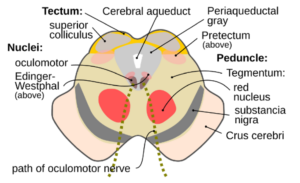 At the most severe end of the spectrum, dysregulation extends into the brainstem structures responsible for autonomic integration itself. Autonomic regulation begins to fragment at its source (Porges, 2011). Cardiovascular regulation, respiration, swallowing, digestion, vestibular function, and cranial nerve output no longer function as a coordinated whole. The clinical picture reflects failure of brainstem integration rather than dysfunction of an isolated organ.
This section sits within an active scientific debate about how persistent trauma-related and autonomic dysregulation should be understood. Somatic trauma models emphasize the ongoing bodily expression of threat. Predictive-processing and active-inference models emphasize persistent predictions of danger and the ways the nervous system weights incoming interoceptive and sensory information. Circuit-based models focus on structures such as the PAG, hypothalamus, amygdala, paraventricular thalamus, habenula, medullary nuclei, and autonomic pathways that participate in selecting and maintaining defensive states. These frameworks are not necessarily competing explanations. They may describe different levels of the same distributed survival system: subjective experience, prediction and interpretation, neural-circuit organization, and bodily physiology.
In simpler terms, researchers may be describing the same elephant from different sides. One group studies how trauma feels and appears in the body, including tension, pain, numbness, panic, digestive changes, collapse, poor sleep, or difficulty moving forward. Another studies how the brain learns to expect danger and continues predicting danger after the original threat has passed. Another studies the brain circuits that select whether the body fights, flees, freezes, shuts down, rests, digests, or connects. Others study the hormones, immune signals, gut, heart, lungs, and muscles that carry out those instructions. No single group has the whole answer. Each is examining one part of a continuously communicating survival system.
The body does not have to store trauma like a file cabinet for trauma to have lasting bodily consequences. Repeated threat can train the brain and body to keep expecting danger. The heart, gut, muscles, immune system, sleep system, hormones, and sensory systems can then send signals that reinforce that expectation. The loop may continue long after the original danger has ended. For this reason, changing one part of the loop may be helpful without being sufficient to reorganize the entire pattern. This does not mean the body is incidental or merely downstream. Signals from the heart, lungs, gut, muscles, immune system, and sensory pathways continuously shape the brain’s predictions and defensive responses.
Within this broader network, the following section considers PAG-mediated tonic immobility as one possible organizing mechanism in the most severe presentations of autonomic dysregulation.
PAG-Mediated Tonic Immobility
While this presentation reads clinically as advanced autonomic fragmentation, this synthesis proposes that its underlying mechanism may be an active, subcortical survival state: periaqueductal gray (PAG)-driven tonic immobility (Lanius et al., 2010; Terpou et al., 2019).
Rather than a simple passive decay of brainstem nuclei, this state represents a subcortical “biological lock-up.” The PAG serves as a primary midbrain command center for survival defenses. When active fight-or-flight (sympathetic dominance) fails or becomes physically impossible, the PAG recruits downstream medullary circuits, including the nucleus tractus solitarius (NTS), dorsal motor nucleus of the vagus (DMNX), and rostral ventrolateral medulla (RVLM), to enforce a high-tone freeze (Porges, 2011; Terpou et al., 2019). This creates a high-energy state where intense sympathetic outflow to the core coexists simultaneously with profound dorsal vagal activation, effectively driving with locked brakes (Levine, 2010).
These presentations appear most frequently under severe physiological or developmental stress where total threat load exceeds the system’s capacity to maintain homeostatic buffering (McEwen, 2007):
- Catastrophic Interoceptive & Surgical Shock: Bone marrow replacements, open heart surgery, end-stage chemotherapy, solid organ transplantation, and major musculoskeletal arthroplasty (such as total joint replacement). The combination of massive mechanical trauma, bone reaming, surgical denervation, acute fluid shifts, and sudden loss of familiar proprioceptive feedback floods the spinothalamic tracts (Barral & Croibier, 2009; Langevin, 2006). In this model, the PAG interprets this abrupt loss of internal structural integrity as a total physical breach, pulling the emergency brake to enforce systemic freeze.
- Persistent Neuroinflammation: Severe Long COVID, where persistent vascular inflammation, microglial activation, and spike protein pathology may affect brainstem nuclei, perpetuating an unceasing state of subcortical threat perception.
- Sensitized Threat Thresholds (DTD): Advanced developmental trauma with deep repression (van der Kolk, 2005; Heller & LaPierre, 2012). Early-life threat “kindles” the PAG, drastically lowering its activation threshold (Terpou et al., 2019; Heim et al., 2008). In these systems, a major physical insult or surgical procedure immediately trips the midbrain emergency brake, bypassing conscious emotional or cortical processing before conscious processing can intervene. (van der Kolk, 2014).
While the manifestations differ, they reflect the same underlying problem: loss of coherent communication within the autonomic system itself driven by an emergency midbrain lockdown. Because this high-tone immobility state is governed by pre-conscious subcortical structures, top-down cognitive or conversational interventions are completely ineffective (van der Kolk, 2014; Schore, 2012). Reversing this lock-up requires bottom-up somatic strategies designed to alter extracellular matrix density (Langevin, 2006; Schleip, 2003), restore afferent vagal signaling to the NTS (Porges, 2011), and modify intrathoracic pressure gradients to directly signal physiological safety back to the PAG (McCraty et al., 2009; Levine, 2010).
Characteristic findings include:
- Advanced cranial nerve dysfunction
- Bradycardia (BradyPOTS) and severe orthostatic intolerance
- Baroreceptor dysfunction
- Central sleep apnea
- Dysphagia and dysphonia
- Severe cardiovascular dysregulation
- Respiratory dysregulation
- Gastroparesis
- Central spasticity and high-tone somatic bracing (Masgutova & Akhmatova, 2011)
This state is not necessarily expressed uniformly across the autonomic system. Developmental history, the timing and nature of threat, and the individual’s subjective experience of that threat can shape which regulatory systems become most strongly conditioned (van der Kolk, 2005; Heim et al., 2008). One person may show profound motor and cranial nerve dysfunction while retaining relatively intact endocrine regulation; another may show severe cardiovascular or gastrointestinal dysregulation with less obvious motor impairment. The same individual may also move between different configurations over time. The underlying pattern is therefore not an all-or-nothing state, but a variable organization of autonomic, endocrine, metabolic, and motor systems around a persistent threat response (McEwen, 2007).
When tonic immobility becomes chronic, the physiological cost can extend beyond the immediate freeze response. Persistent sympathetic activation can coexist with suppressed HPA activity, producing the paradoxical combination of high noradrenergic drive and low baseline cortisol (Heim et al., 2008; Yehuda et al., 2016).
Some Consequences of Chronic Tonic Immobility
The consequences of chronic tonic immobility extend beyond the immediate autonomic state because the systems involved in maintaining physiological stability do not operate independently. Persistent vlPAG activation can maintain GABAergic and glycinergic inhibition of spinal motor output while sympathetic and noradrenergic circuits remain active (Lanius et al., 2010; Terpou et al., 2019). At the same time, dorsal vagal activation alters gastrointestinal motility, secretion, and visceral signaling (Porges, 2011). The resulting state is not simply a brainstem problem. It is a distributed physiological configuration in which central, autonomic, enteric, endocrine, immune, metabolic, and sensory systems continuously influence one another (McEwen, 2007).
The metabolic consequences illustrate this interdependence. When automatic motor organization through the basal ganglia is disrupted by persistent defensive inhibition, the medial and dorsolateral prefrontal cortices (mPFC and dlPFC) may have to supply conscious, step-by-step control for actions that would ordinarily occur with little cortical effort. This substantially increases executive and metabolic demand. At the same time, chronic HPA axis suppression can produce low baseline cortisol, reducing the metabolic and anti-inflammatory support normally available to the system (Chapman et al., 2013; Heim et al., 2008). Persistent locus coeruleus activity can maintain high noradrenergic output even while the HPA axis remains blunted. The result can be profound exhaustion without a corresponding reduction in internal autonomic activation.
Dopamine is one component of this larger network. Cortisol supports the biochemical environment required for catecholamine synthesis, including tyrosine hydroxylase (TH) (Chapman et al., 2013), while persistent noradrenergic demand places continued pressure on catecholamine resources. A system with reduced cortisol support and persistent locus coeruleus activity may therefore have difficulty sustaining dopamine-dependent executive function, motor initiation, reward processing, and goal-directed behavior (McEwen, 2007). This provides one mechanism through which a person can remain highly activated internally while experiencing cognitive fog, impaired initiation, reduced motor fluency, anhedonia, or profound fatigue. These findings cannot, however, be reduced to a dopamine deficit because the same defensive state is simultaneously altering multiple other regulatory systems.
The gastrointestinal system is one of those systems. The enteric nervous system, vagal pathways, intestinal immune system, and gut microbial environment all participate in signaling the physiological state of the body back to the brain (Davis, 2022; Porges, 2011). Persistent sympathetic activation and dorsal vagal dominance can alter motility, secretion, circulation, and gut barrier function. When cortisol regulation is chronically impaired, the loss of cortisol’s normal effects on epithelial integrity and immune regulation can further increase vulnerability to intestinal barrier disruption (Chapman et al., 2013). Increased permeability then permits greater interaction between luminal contents, immune cells, and the systemic circulation, creating another source of inflammatory signaling rather than the stable visceral environment associated with physiological safety (Davis, 2022).
The consequences extend into gut neurochemistry and immune signaling. Altered enteric function changes the peripheral environment in which serotonin and other signaling molecules are regulated, while changes in the microbial ecosystem can alter production of microbial metabolites, including short-chain fatty acids (SCFAs), that participate in communication among the gut, immune system, and nervous system (Davis, 2022). Increased intestinal permeability can also increase exposure to microbial products that activate innate immune pathways and cytokine signaling. These signals can influence vagal afferents, brainstem nuclei, microglial activity, and central inflammatory signaling (Porges, 2011; Moffitt, 2025). Thus the gut is not simply an organ affected by autonomic dysregulation. It can become an active source of signals that reinforce the very state of dysregulation that impaired its function.
The immune system adds another layer to the same network. Low cortisol removes part of the endogenous restraint on inflammatory activity (Chapman et al., 2013), while persistent sympathetic signaling directly affects immune tissues (McEwen, 2007). Cytokine signaling can in turn alter central nervous system function, energy availability, behavior, and interoceptive processing. The resulting feedback does not move in a single direction. Brainstem activity affects the gut and immune system; gut and immune signals return through visceral and humoral pathways; endocrine changes alter both systems; and the resulting signals feed back into central autonomic and defensive circuits (McEwen, 2007).
Endogenous opioid signaling provides another example. Persistent vlPAG activation can recruit -endorphins and dynorphins, producing analgesia and altered conscious experience during extreme threat (Lanius et al., 2010; Terpou et al., 2019). When maintained chronically, these mechanisms may contribute to emotional blunting, dissociation, cognitive impairment, and altered reward processing (van der Kolk, 2014). At the same time, endocrine changes can affect thyroid metabolism and reproductive signaling (Kalantaridou et al., 2004), while immune and inflammatory changes can further alter energy availability and central processing. Each system therefore becomes both an effect of the defensive state and a potential source of feedback that maintains it.
This is why the clinical expression cannot be reduced to a single neurotransmitter, hormone, organ, or receptor. Dopamine, serotonin, cortisol, norepinephrine, GABA, endogenous opioids, cytokines, gut-derived metabolites, vagal signaling, and cortical and brainstem circuits exist within a continuously interacting system. Altering one component may change one part of the physiology without changing the organization of the network that produced the dysregulation. A pharmacological intervention directed at a single molecule may therefore produce a limited, transient, or poorly tolerated effect when the surrounding physiological conditions continue to generate the same defensive state (McEwen, 2007; van der Kolk, 2014).
This network is also not expressed uniformly. Developmental history, the timing and nature of threat, and the individual’s subjective experience determine which circuits become most strongly conditioned (van der Kolk, 2005; Heim et al., 2008). One person may develop prominent cranial nerve and motor manifestations, another severe gastrointestinal or immune dysfunction, another marked endocrine and metabolic disruption, and another substantial cognitive or reward system impairment. Several systems may remain relatively intact while others become profoundly dysregulated. The clinical picture therefore reflects the particular configuration of the network in that individual rather than a single biochemical signature of tonic immobility.
Hub: The Autonomic Nervous System – The Corrupted Blueprint
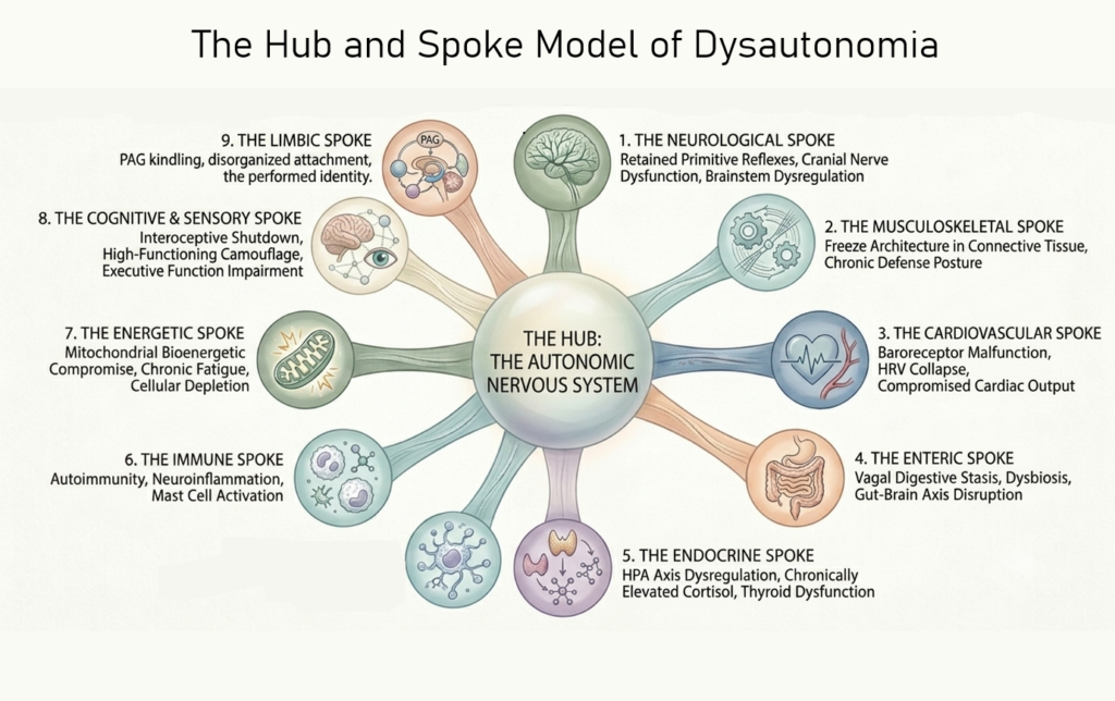
The organizing principle here is built around a hub and spoke model. The hub is the autonomic nervous system, whose operating system has been corrupted by chronic dysregulation. The spokes are the distinct systems where that corruption shows up as observable pathology.
The spokes are not isolated. Dysautonomia patients present with multiple systems involved, and understanding which combination is active determines both the clinical picture and where treatment begins. The same patient may accumulate neurological, psychiatric, gastrointestinal, autoimmune, and developmental diagnoses without anyone identifying the autonomic pattern that links them. Developmental trauma patients show this layering at its most severe, often carrying deficits across every system at once.
- The neurological spoke: retained primitive reflexes, cranial nerve dysfunction, and demyelination.
- The musculoskeletal, fascial spoke: the freeze architecture written into the connective tissue and the sinew channel expression of chronic defense.
- The cardiovascular spoke: baroreceptor malfunction and HRV collapse.
- The enteric spoke: vagal driven digestive stasis, dysbiosis, and gut brain axis disruption.
- The endocrine spoke: HPA axis dysregulation, thyroid dysfunction, and disrupted steroidogenesis.
- The immune spoke: autoimmunity, neuroinflammation, and mast cell activation.
- The energetic spoke: mitochondrial compromise, cellular bioenergetic depletion, and the heritable deficit.
- The cognitive and sensory spoke: the developmental wiring cluster, interoceptive shutdown, and the high functioning camouflage.
- The limbic spoke: PAG kindling, disorganized attachment architecture, and a self that learned to perform instead of exist.
These are not separate diagnoses requiring separate specialists. They are the predictable downstream consequences of one central operating system forced to run on a corrupted blueprint for a lifetime. In classical TCM, treating the branch without the root produces temporary relief at best. The same principle applies here: a clinician who treats only the spoke never reaches the hub, and the patient never gets better.
Once you recognize the pattern in one spoke, the clinical question expands: where else does it present? Every element introduced here is explored in the sections that follow. The patterns described here are by no means complete. They represent what has been mapped thus far. As more practitioners across modalities begin working with these principles, the patterns will continue to expand.
The spokes are presented separately for clarity, but they neither develop nor function in isolation. They develop together as an integrated system during prenatal development and childhood and remain physiologically interconnected throughout life. This principle becomes important in the treatment discussion, where access through a single system can produce change across multiple systems simultaneously.
Developmental Trauma Disorder: The Worst Case
Every spoke in this framework can appear without a history of childhood trauma. Chronic pain, autoimmune disease, long COVID, and neurodegenerative disease all produce the same autonomic patterns described here. But Developmental Trauma Disorder represents the most severe and treatment resistant expression of this picture, because the autonomic dysfunction did not happen to an already formed nervous system. It happened during the years that system was still developing. The hub was corrupted before it finished development, not damaged after the fact, so most, if not all, spokes carry some degree of impairment.
 Continued here…