Spoke 1: The Neurological Spoke — Cranial Nerve Involvement and Brainstem Dysregulation
Dysautonomia is fundamentally a disorder of autonomic regulation, making the brainstem the logical starting point for clinical assessment. (Porges, 2011)
Visual pathways carry more than image-forming information. Retinal signals participate in circadian timing, hypothalamic regulation, orientation, and autonomic state selection. In patients with developmental trauma, head injury, or persistent visual-field disturbance, altered sensory gating may contribute to ongoing autonomic load. A detailed discussion of visual processing, thalamic gating, and mitochondrial implications is provided separately.
Cranial nerve involvement in the clinical examination indicates that the brainstem is in chronic high arousal. Facial asymmetry, dysphagia, voice changes, eye tracking deficits, jaw locking, palatal collapse, tongue deviation, hyperacusis, and the constellation of vagal symptoms: gastroparesis, bradycardia or tachycardia, and orthostatic intolerance are not findings about isolated cranial nerve function. They are findings about the autonomic state of the brainstem nuclei from which those cranial nerves originate. The problem is not in the nerve; it is in the brainstem. (Pryor, 2019)
Retained primitive reflexes provide another window into the same process. Primitive reflexes such as the Moro, Fear Paralysis Reflex (FPR), and Spinal Galant can remain reactive when normal developmental integration is disrupted, or they may reactivate following overwhelming psychological or physiological stress. Masgutova’s MNRI research demonstrated that a significant traumatic event can reactivate previously integrated primitive reflexes, and that they can remain active for months or years following that event. (Masgutova & Akhmatova, 2011) This is the body level evidence of what gets treated as a purely psychological condition in PTSD. The finding is important because persistent primitive reflexes indicate a nervous system in a chronic high state of arousal. Neuroplasticity is diminished, higher order processing is impaired, and the capacity for learning, adaptation, and recovery declines. (McEwen, 2007; Pryor, 2019)
Together, cranial nerve findings and retained primitive reflexes point to a nervous system organized around defense rather than regulation. Several theoretical frameworks describe different aspects of this same phenomenon.
For a fuller discussion of brainstem regulation, cranial nerve function, and related clinical patterns, see “Brainstem Regulation, Cranial Nerve Function, and Sensorimotor Organization.
Spoke 2: The Musculoskeletal and Fascial Spoke: The Freeze Pattern Written into Connective Tissue and the Sinew Channel Expression of Chronic Defense
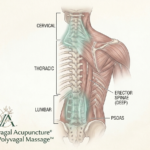
If the neurological spoke describes the origin of dysautonomia, the musculoskeletal system reveals its biological footprint. (Porges, 2011). Chronic autonomic defense is ultimately written into the body’s connective tissue. (Schleip, 2003). The primary musculoskeletal finding across dysautonomia is a chronic protective bracing pattern. (Lowen, 1975). This pattern is centrally mediated through persistent autonomic output from the brainstem rather than arising within the muscle or fascia itself. (McEwen, 2007). Consequently, these areas of rigidity cannot simply be massaged or stretched away because the nervous system continually recreates the pattern until the underlying autonomic state changes. (Levine, 2010).
Spasticity is abnormal muscle stiffness resulting from damage to the upper motor neurons in the brain or spinal cord. That damage disrupts normal regulation of the stretch reflex, causing muscles to remain contracted, and resist passive movement. (Lance, 1980). Dysautonomia can produce a similar clinical presentation without upper motor neuron damage, arising instead from conflicting flexion and extension synergies acting on the tissue. (Levine, 2010). Spasticity, though, is only one of the findings. Tone, color, fascial density, movement, glide, circulation, and tissue texture are the markers. You can see the autonomic state in the tissue itself.
Carla Stecco, an orthopedic surgeon and anatomist, demonstrated that fascia is continuously replaced rather than simply repaired. (Stecco, 2015) Chronic dysautonomia alters that remodeling process. Under persistent autonomic defense, fascial remodeling progressively shifts away from normal elastic restoration. The extracellular matrix becomes increasingly chondroitin dominant, elasticity declines, glide diminishes, and the tissue becomes progressively denser, more adherent, and increasingly resistant to movement. (Langevin, 2006) The fascia becomes cement. Certain regions consistently carry the highest burden of this adaptation. The deep cervical territory, axial brachial plexus corridor, scalenes, SCM, upper trapezius, and cervicothoracic junction become primary sites of chronic bracing across dysautonomia patterns and are particularly pronounced in retained freeze architecture. Once established, the pattern fixes through this axis as spasticity, restricted glide, altered recruitment, and persistent tissue density. Tissue that no longer moves stops feeding the vagus the sensory input it depends on. The vagus is roughly eighty percent afferent, and that afferent traffic tells the brainstem and vestibular system where the body is, what state it is in, and what is happening right now. When fascial movement drops, that feedback drops, and the nervous system reduces engagement with pathways that are no longer consistently activated. Over time, this drives progressive restriction of movement and loss of function through the same use dependent mechanisms that drive synaptic pruning throughout the nervous system. (McEwen, 2007) Much of what we attribute to normal aging may instead reflect the cumulative consequences of diminished movement, reduced sensory input, and chronic autonomic dysregulation. (Erickson et al., 2011)
These tissue level adaptations produce a remarkably consistent clinical picture across disorders traditionally considered unrelated. Patients across the dysautonomia spectrum present with severe high and low tone, including spasticity, stiff person presentations, and MS-type sinew patterns, along with limited range of motion and pain. Areas of chronic contraction and guarding sit beside areas of collapse and poor recruitment. The same picture runs across neurogenic conditions, including Parkinson’s and stroke sequelae, and through cases nobody usually describes this way. PTSD patients carry that density and restricted movement in their necks and shoulders. (Masgutova, 2011). Chronic orthopedic injuries that never resolved develop it too: thick, immobile tissue around the injury site, with weak muscle activation and reduced circulation nearby. It appears wherever autonomic dysregulation has lasted long enough to alter tissue repair. Developmental Trauma Disorder represents the most severe and treatment resistant expression of it. (van der Kolk, 2014)
For clinicians without a neurological background, the fascial system often provides the most accessible window into autonomic dysfunction.These changes are frequently evident on routine physical examination through diminished range of motion, altered posture, and standard soft tissue orthopedic assessment, and they become even more apparent through skilled palpation. (Schleip, 2003). Most psychotherapists are not trained to evaluate these findings, but bodyworkers, fascial therapists, physical therapists, and other clinicians trained in manual assessment can often recognize them immediately. (Moffitt, 2024). This distinction carries important implications for treatment. Many trauma therapies emphasize cognitive processing, emotional regulation, or other top-down strategies. (van der Kolk, 2014). These approaches may reduce distress and improve coping, but they cannot by themselves reverse years of altered fascial remodeling, persistent protective motor patterns, or diminished afferent signaling. (Levine, 2010; Schleip, 2003). When the tissue itself has become part of the pathology, psychotherapy and body-based treatment become complementary rather than competing approaches. (Levine, 2010; van der Kolk, 2014).
As treatment progresses, the tissue changes in ways visible, palpable, and objective to both patient and practitioner, so the same signs that named the problem now confirm the work as it happens. Areas that were fixed begin to soften and move in real time. Color, circulation, and tone shift, and you can document these changes through palpation, photographs, and standard soft tissue orthopedic testing. Fascial glide returns, elasticity restores, areas of hemosiderin deposition and dusky tissue resolve, and normal tissue architecture returns, establishing the objective clinical endpoint of restored vagal patency. (Langevin, 2006; Pryor, 2023).
 Continued here…
AI-assisted drafting was used in the organization of the material.
References
Balaban, C. D., & Thayer, J. F. (2001). Neurological bases for balance-anxiety links. Journal of Anxiety Disorders, 15(1–2), 53–79.
Chan, A., Harvey, P., Hernandez-Cardenache, R., Alperin, N., Lee, S., Hunt, C., Petersen, N., Northoff, G., Robertson, N., Ouyang, J., Karasik, R., & Williams, K. (2024). Trauma and the default mode network: Review and exploratory study. Frontiers in Behavioral Neuroscience, 18, 1499408.
Damasio, A. R. (1994). Descartes’ error: Emotion, reason, and the human brain. Putnam.
Deadman, P., & Al-Khafaji, M. (2007). A manual of acupuncture (2nd ed.). Journal of Chinese Medicine Publications.
Goyal, M. S., Hawrylycz, M., Miller, J. A., Snyder, A. Z., & Raichle, M. E. (2014). Aerobic glycolysis in the human brain and its implications for cellular and vascular biology. Neuron, 81(2), 289–296.
Heller, L., & LaPierre, A. (2012). Healing developmental trauma: How early trauma affects self-regulation, self-image, and the capacity for relationship. North Atlantic Books. [1]
Hull, L., Petrides, K. V., Allison, C., Smith, P., Baron-Cohen, S., Lai, M. C., & Mandy, W. (2017). Putting on my best normal: Social camouflaging in adults with autism spectrum conditions. Journal of Autism and Developmental Disorders, 47(8), 2519–2534.
Ilveskoski, I., Lehtimäki, K., & Mäkinen, J. (1993). Myelination of vestibular pathways in the developing human brain. Acta Oto-Laryngologica, 113(2), 178–182.
Kandel, E. R., Koester, J. D., Mack, S. H., & Siegelbaum, S. A. (2021). Principles of neural science (6th ed.). McGraw-Hill.
Karnath, H. O. (2007). Spatial orientation and the representation of space with parietal cortex. Philosophical Transactions of the Royal Society B: Biological Sciences, 362(1480), 1411–1421.
LeDoux, J. E. (1996). The emotional brain: The mysterious underpinnings of emotional life. Simon and Schuster.
Levine, P. A. (2010). In an unspoken voice: How the body releases trauma and restores goodness. North Atlantic Books.
Llewellyn-Smith, I. J., & Verberne, A. J. M. (Eds.). (2011). Central regulation of autonomic functions (2nd ed.). Oxford University Press.
Masgutova, S., & Akhmatova, N. (2011). Integration of dynamic and postural reflexes into the whole body movement system. MNRI Method.
Maté, G. (2019). When the body says no: The cost of hidden stress. Vintage Canada.
McDonnell, M. N., & Hillier, S. L. (2015). Vestibular rehabilitation for unilateral peripheral vestibular dysfunction. Cochrane Database of Systematic Reviews, 1, CD005397.
Moffitt, J. (2024). Primitive reflexes and their role in neurodevelopment and emotional regulation. Down the Polyvagal Rabbit Hole. Polyvagal Acupuncture®.
Moffitt, J. (2025a). The serotonin-disgust-attachment suppression mechanism in DTD. Polyvagal Acupuncture®.
Moffitt, J. (2025b). The great doubt and the return of self-initiated presence. Polyvagal Acupuncture®.
Myers, T. W. (2020). Anatomy trains: Myofascial meridians for manual and movement therapists. Churchill Livingstone.
Panksepp, J. (1998). Affective neuroscience: The foundations of human and animal emotions. Oxford University Press.
Porges, S. W. (2011). The polyvagal theory: Neurophysiological foundations of emotions, attachment, communication, and self-regulation. Norton.
Raichle, M. E., & Gusnard, D. A. (2002). Appraising the brain’s energy budget. Proceedings of the National Academy of Sciences, 99(16), 10237–10239.
Schore, A. N. (2012). The science of the art of psychotherapy. Norton.
Spear, A. D. (2019). Epistemic dimensions of gaslighting: Peer-disagreement, self-trust, and epistemic injustice. Inquiry, 66(1), 11–36.
Stein, B. E., & Meredith, M. A. (1993). The merging of the senses. MIT Press.
Tamietto, M., & de Gelder, B. (2010). Neural bases of the non-conscious perception of emotional signals. Nature Reviews Neuroscience, 11(10), 697–709.
van der Kolk, B. A. (2014). The body keeps the score: Brain, mind, and body in the healing of trauma. Viking.
Winnicott, D. W. (1965). The maturational processes and the facilitating environment: Studies in the theory of emotional development. International Universities Press
Yehuda, R., & Lehrner, A. (2018). Intergenerational transmission of trauma effects: Putative role of epigenetic mechanisms. World Psychiatry, 17(3), 243–257.
Yuksel, E., Mukherjee, M., & Amiri, M. (2020). Is forward head posture relevant to autonomic nervous system function and cervical sensorimotor control? Gait and Posture, 77, 71–74.