This synthesis emerged during COVID after more than 25 years of clinical practice and my own experience of autonomic collapse from developmental trauma. The mechanistic reasoning is grounded in neuroanatomy, autonomic physiology, mitochondrial bioenergetics, and developmental neuropsychology. The clinical frameworks, techniques, and synthesis presented in this series reflect an approach that has been developed and applied in clinical practice over the last several years, largely to facilitate my own neuroplastic recovery.. The opinions expressed here are mine.
This article introduces protective bracing as a way to recognize how a body remains organized around protection. I use the term protective bracing to refer to chronic fascial patterns in the body and how these systems reflect a nervous system in a high state of arousal. It can present as sustained contraction, restricted movement, altered breathing, gait changes, endocrine imbalance, metabolic dysfunction or chronic muscle tightness or rigidity.
The purpose is not to reduce bracing patterns to posture, fascial restriction, or a specific reflex. Rather, the goal is to identify observable protective patterns in the fascia to facilitate their release gradually, from the body upward.
Clients usually enter treatment because of pain, exhaustion, sleep disturbance, cognitive changes, or movement restriction. The clinical task is to recognize when those concerns reflect a body still held in a protective response after the initiating threat has passed. This article develops protective bracing as a clinical framework to begin to restore balance to the nervous system.
This work emerged from severe presentations of dysautonomia, particularly in Long COVID and severe neurogenic conditions that include TBI and developmental trauma. Although these conditions present differently, the autonomic nervous system and its balance emerged as a central point of assessment and treatment, alongside the restoration of neuroplastic function. The focus became the restoration of autonomic balance in a nervous system that had become persistently dysregulated. The fascia provides a clinical entry point for recognizing and working with this retained protective organization, and these patterns provide a way to see where the body is still holding a defensive response.
Protective bracing should not automatically be equated with neurologically defined spasticity. In conventional neurology, spasticity refers specifically to velocity-dependent increased muscle tone associated with exaggerated stretch reflexes and, classically, upper motor neuron dysfunction (Sheean & McGuire, 2009). A person may have neurological spasticity and also show a broader bracing pattern. Others may show rigidity, guarding, sustained co-contraction, or postural fixation without a documented upper motor neuron lesion. This model distinguishes neurological spasticity from other forms of guarding and bracing, although each reflects persistent protective organization. This distinction creates a gateway for somatic practice through skilled practitioner intervention and patient-directed daily practices that restore orientation, interoception, mobility, respiration, and tolerance for safe movement. The aim is not to force release or infer a lesion or single retained reflex from posture alone, but to identify what maintains the pattern and provide graded conditions in which flexibility and regulation become possible (Levine, 2010).
Sinew Channels, Fascial Lines, and Protective Bracing
The sinew channels, or jing jin, are one of the channel systems of Traditional Chinese Medicine. They are distinct from the primary channels and provide the framework used in this work to examine protective bracing in the body.
Reproduced with kind permission of amanualofacupuncture.com’.
In TCM, the twelve primary meridians are organized into six divisions: Tai Yang, Yang Ming, Shao Yang, Tai Yin, Shao Yin, and Jue Yin. Each division includes paired arm and leg channels. The six divisions became particularly important as a framework for understanding the penetration and progression of disease within the herbal medical system described in the Shang Han Lun and related texts. The classical jing jin are also organized through these same six divisions, rather than being treated as twelve separate meridians.
The classical sinew channels provide a full body map for observing how protection is somatized and retained as protective bracing in the body. They link the limbs, spine, cranium, posture, movement, emotions, and autonomic response rather than limiting assessment to isolated muscles or joint restrictions. For the purpose of examining patterns of protective bracing and retained threat responses, this whole body organization provides a useful framework for understanding how protective patterns are expressed through fascia. During development, the autonomic nervous system develops alongside the gut, immune, limbic, and musculoskeletal systems. This shared development is one reason fascia provides an effective route through which we can begin to restore balance to the nervous system.
Thomas Myers’ work on Anatomy Trains provides modern myofascial language for understanding fascial continuity and the relationships described through the sinew channels. His model describes how tension, biomechanical load, and restricted movement often extend beyond the local site where symptoms appear. The posterior, anterior, lateral, spiral, and deep front lines make visible forms of continuity that are often clinically evident but easily missed when symptoms are treated in isolation (Myers, 2020).
Alongside Myers, Luigi and Carla Stecco’s work on the structure, function, and innervation of fascia provides contemporary anatomical language for the tissue systems involved. The work of Svetlana Masgutova, Peter Levine, Bessel van der Kolk, and other somatic and trauma researchers has described in detail how developmental and threat responses become expressed through autonomic state, movement, posture, respiration, and bodily organization. Physical therapy and neurology provide the clinical markers that make it possible to objectively follow changes in autonomic balance, vagal tone, and neuroplastic function.
In this approach, we use “sinew channels” and “myofascial lines” interchangeably. Although they are not one to one anatomical equivalents, using both terms allows clearer communication across modalities through a shared clinical language. TCM practitioners can work through the deeper sinew channel system, while other clinicians can describe the same whole-body fight, flight, or freeze pattern through myofascial lines.
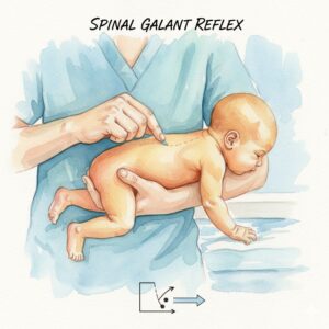
Primitive reflexes are brainstem-mediated survival responses that organize early orientation and postural development. Their emergence is directed through cranial-nerve development, which organizes feeding, sensory orientation, facial and head movement, airway function, and the infant’s early relationship to the environment.
As cranial nerve function develops, primitive reflexes do not remain separate motor events. Postural reflexes become increasingly integrated with vestibular balance and movement. Reflexive orientation and protection become integrated with limbic or emotional response. Later, these systems come under the influence of higher processing, cognition, voluntary movement, gait, and autonomic regulation.
When early patterns remain active, reactivate, or are recruited under stress, they reflect a nervous system in a high state of arousal. This state can keep the nervous system locked in protection, making it harder for the body to settle, reorganize movement, and repair through neuroplastic change. In adulthood, these patterns rarely appear as textbook infant reflexes. They may instead be recognized through sinew rigidity, asymmetrical tension, restricted breathing, and loss of reciprocal movement.
Whole-Body Patterning
Protective bracing may appear in a localized region while expressing a larger pattern. Upper-cervical restriction may involve the posterior chain, suboccipital tissues, jaw, hyoid, anterior neck, clavicular triangle, thoracic inlet, scapular organization, diaphragm, and pelvic loading. A lateral pattern may involve the Gallbladder sinew channel, lateral fascial line, rib cage, pelvis, shoulder, and cervical side-bending. A deep freeze pattern may involve the internal rotators, pelvic floor, diaphragm, deep anterior structures, jaw, and airway organization.
The clinical question is not only where tissue feels restricted. It is how the body has organized the restriction and what protective function that organization may still be serving. How the body organizes its response to threat may reveal patterns that earlier clinicians recognized in practice, even before modern science could explain them
Hinge Zones
A hinge zone is a region where several protective patterns meet through shared structural and autonomic pathways. A hinge zone is not the root cause of a problem, but rather a useful starting point for understanding and addressing protective patterns in the body. These zones are clinically important because they represent points where multiple layers of fascia and defensive systems converge. Intervening here can influence several layers and functions at once.
The upper cervical region is one of the most significant hinge zones. The occiput, C1–C2, suboccipitals, jaw, hyoid, tongue, anterior neck, clavicular triangle, upper ribs, and thoracic inlet bring together cranial protection, head orientation, postural control, airway and swallowing organization, voice, cervical movement, and brainstem-related signaling. A sustained bracing pattern in this area affects far more than neck range of motion (Lanius et al., 2014).
Other hinge zones include the thoracolumbar junction, diaphragm and lower ribs, pelvic floor and sacrum, shoulder girdle and upper ribs, and the pelvis-lower-extremity relationship. A pattern may be expressed most visibly in one area while maintained through another. For example, cervical bracing may be reinforced by restrictions elsewhere in the body, particularly through the thoracic inlet, diaphragm, or pelvis.
Clinical Application of the Model
Once a dysfunctional pattern is identified, the clinician selects an intervention entry point based on the individual’s current autonomic state and tolerance. Interventions may start with manual contact, movement, breathwork, or sinew-channel techniques. The same graded approach informs patient self-practice between sessions to encourage manageable adjustments in movement, breathing, and tension that support ongoing neuroplastic repair.
The therapeutic objective is to recognize cues of a retained fight-or-flight response and help the body return to a more balanced state of autonomic regulation. (Levine, 2010).
Observable Markers of Vagal Tone Restoration
As bracing lessens, the body shows smoother fascial movement, improved reciprocal motion, fuller breathing, reduced co-contraction, better orientation, and greater presence during activity. Regional color change that occurs with these shifts, particularly in somatic dysautonomia, is clinically relevant and indicates objective restoration of vagal tone.
Toward PAG Cervical Bracing
One upper-cervical pattern has become especially important in my own neuroplastic process and in work with complex trauma physiology: a PAG Cervical Bracing Pattern. This configuration involves the occiput, C1–C2, jaw-hyoid complex, anterior neck, clavicular triangle, upper thoracic inlet, and related cranial, respiratory, postural, and brainstem-linked protective organization.
The next article examines this pattern as a working neurodevelopmental and neurosomatic model. It considers how overlapping protective reflex patterns, chronic trauma physiology, cranial restriction, fascial fixation, respiratory and jaw-hyoid dysfunction, and possible brainstem involvement may converge into persistent adult cervical bracing.
References
Lanius, R. A., Paulsen, S. L., & Corrigan, F. M. (Eds.). (2014). Neurobiology and treatment of dissociation: Toward an embodied self. Springer. https://doi.org/10.1007/978-1-4614-6086-9
Levine, P. A. (2010). In an unspoken voice: How the body releases trauma and restores goodness. North Atlantic Books.
Moffitt, J. (2026). The Chong Mai: An integrative neurological framework. Polyvagal Acupuncture®.
Myers, T. W. (2020). Anatomy trains: Myofascial meridians for manual and movement therapists (4th ed.). Elsevier.
Sheean, G., & McGuire, J. R. (2009). Spastic hypertonia and movement disorders: Pathophysiology, clinical presentation, and quantification. PM&R, 1(9), 827-833. https://doi.org/10.1016/j.pmrj.2009.08.002
UCLA Health. (2023, January 2). Hemosiderin staining: Product of trauma or venous insufficiency. UCLA Health.
van der Kolk, B. A. (2014). The body keeps the score: Brain, mind, and body in the healing of trauma. Viking.
Yu, S. K., Kim, T. H., Yang, K. Y., Bae, C. J., & Kim, H. J. (2021). Morphology of the temporalis muscle focusing on the tendinous attachment onto the coronoid process. Anatomy & Cell Biology, 54(3), 308-314. https://doi.org/10.5115/acb.21.074


 As cranial nerve function develops, primitive reflexes do not remain separate motor events. Postural reflexes become increasingly integrated with vestibular balance and movement. Reflexive orientation and protection become integrated with limbic or emotional response. Later, these systems come under the influence of higher processing, cognition, voluntary movement, gait, and autonomic regulation.